
The femoral triangle is a hollow area in the anterior thigh. Many large neurovascular structures pass through this area, and can be accessed relatively easily. Thus, it is an area of both anatomical and clinical importance.
In this article, we shall look at the borders, contents and clinical correlations of the femoral triangle.
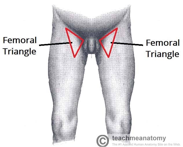
Borders
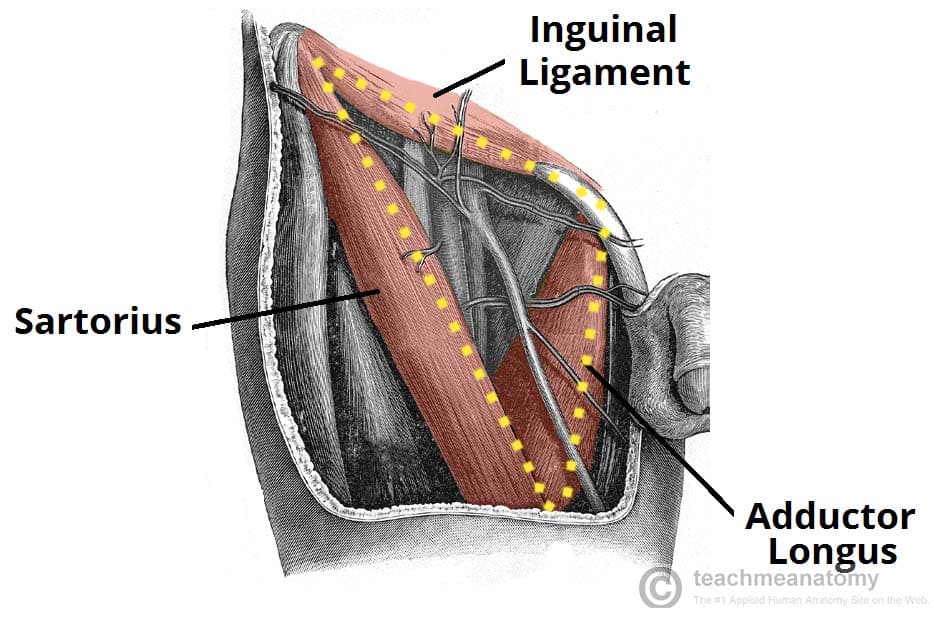
As this area is a triangle, it has three borders:
- Superior border – Formed by the inguinal ligament, a ligament that runs from the anterior superior iliac spine to the pubic tubercle.
- Lateral border – Formed by the medial border of the sartorius muscle.
-
Medial border – Formed by the medial border of the adductor longus muscle. The rest of this muscle forms part of the floor of the triangle.
- Note: Some sources consider the lateral border of the adductor longus to be the medial border of the femoral triangle. However, the majority state that it is the medial border of the adductor longus – and this is definition we have gone with.
It also has a floor and a roof:
- Anteriorly, the roof of the femoral triangle is formed by the fascia lata.
- Posteriorly, the base of the femoral triangle is formed by the pectineus, iliopsoas and adductor longus muscles.
The inguinal ligament acts as a flexor retinaculum, supporting the contents of the femoral triangle during flexion at the hip.
Contents
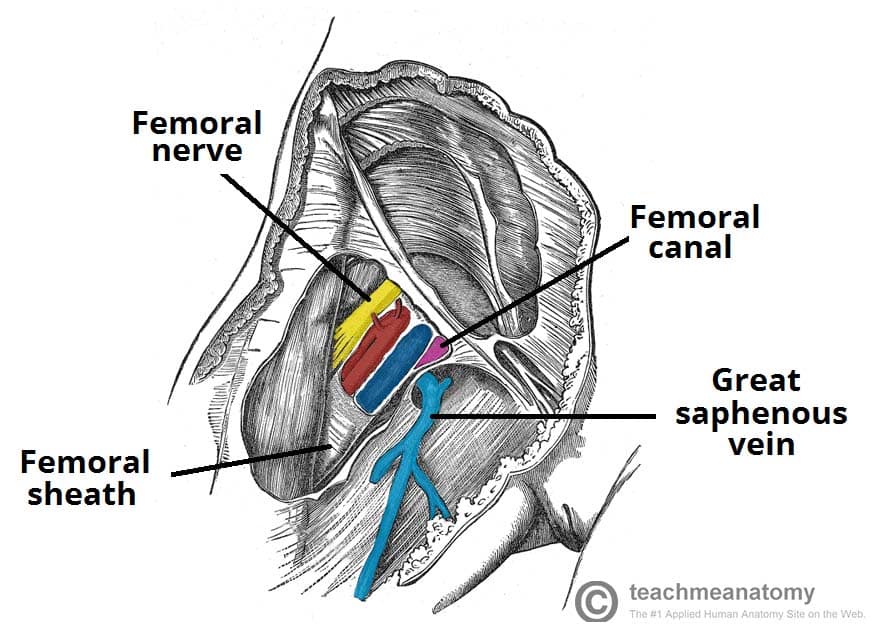
The femoral triangle contains some of the major neurovascular structures of the lower limb. Its contents (lateral to medial) are:
- Femoral nerve – Innervates the anterior compartment of the thigh, and provides sensory branches for the leg and foot.
- Femoral artery – Responsible for the majority of the arterial supply to the lower limb.
- Femoral vein – The great saphenous vein drains into the femoral vein within the triangle.
- Femoral canal – A structure which contains deep lymph nodes and vessels.
The femoral artery, vein and canal are contained within a fascial compartment – known as the femoral sheath.
A good way of remembering the contents is using the acronym NAVEL:
N: Nerve.
A: Artery.
V: Vein.
E: Empty space (this is important as it allows the veins and lymph vessels to distend, so they can cope with different levels of flow).
L: Lymph canal.
Clinical Relevance: The Femoral Triangle
Femoral Pulse
Just inferior to where the femoral artery crosses the inguinal ligament, it can be palpated to measure the femoral pulse. The femoral artery crosses exactly midway between the pubic symphysis and anterior superior iliac spine (known as the mid-inguinal point).
Access to the Femoral Artery
The femoral artery is located superficially within the femoral triangle, and is thus easy to access. This makes it suitable for a range of clinical procedures.
One such procedure is coronary angiography. Here, the femoral artery is catheterised with a long, thin tube. This tube is navigated up the external iliac artery, common iliac artery, aorta, and into the coronary vessels. A radio-opaque dye is then injected into the coronary vessels, and any wall thickening or blockages can be visualised via x-ray.
Femoral Hernia
A hernia is defined as “a condition in which part of an organ is displaced and protrudes through the wall of the cavity containing it“.
In the case of femoral hernia, part of the bowel pushes into the femoral canal, underneath the inguinal ligament.
This manifests clinically as a lump or bulge in the area of the femoral triangle. It usually requires surgical intervention to treat.
